10x Genomics still leads the spatial transcriptomics field with continuous innovations. Among its products, Visium HD and Xenium has introduced a5000-gene panel, optimizing imaging-based gene detection.
Choosing the right platform for your research can be challenging. This article compares Visium HD and Xeniumto help you determine the best fit based on your study objectives, sample type, and budget.

High-Resolution Detection Image Example (Left: Visium HD, Right: Xenium)
Both platforms use probe hybridizationto detect gene expression in tissue sections, but they differ in methodology and data output.
Visium HD: Captures RNA in situusing probes hybridized to a spatially barcoded slide. This allows high-throughput sequencing to generatespatially resolved gene expression matrices.
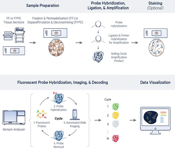
Xenium: Uses fluorescent in situ hybridization (FISH), where rolling circle amplification enhances signals. Multiple rounds offluorescence imaging and decodingon Xenium instruments provide gene expression data with high spatial precision.
Key Differences:
Visium HD is sequencing-based, offering broad transcriptomic coverage.
Xeniumuses imaging, enabling faster and more targeted gene detection.

Visium HD Workflow Diagram

Xenium Workflow Diagram
Both platforms rely on probe-based RNA capture, but their probe designs and applications differ significantly.
Probe Structure:
Visium HD: Probes hybridize to RNA and are captureddirectly on the slide.
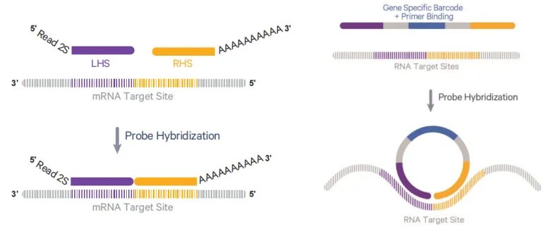
Xenium: Uses a padlock probethat circularizes after hybridization, enablingrolling circle amplification.
Probe Quantity Per Gene:
Visium HD: Most genes are detected usingthree probes per gene.
Xenium: Uses 5–8 probes per gene (with most genes utilizing 8 probes), ensuring higher specificity and sensitivity.

Probe Structure Diagram
(Left: Visium HD Probe Structure; Right: Xenium Probe Structure)
Gene Coverage:
Visium HD: Detects ~18,000 genes (human) and ~19,000 genes (mouse) through sequencing.
Xenium: Detects up to 5000 genes, constrained by fluorescence imaging limitations.
Customization:
Visium HD : Limited to human and mouse.
Xenium:Supports custom probe design for non-human/mouse species, allowing researchers to tailor probe selection based on tissue characteristics.
The slide design reflects differences in detection methods.
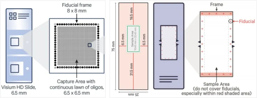
Visium HD: Two sample frames, each 6.5mm × 6.5mm, covered with 2μm × 2μm spatially barcoded grids.
Xenium: One large sample frame (max tissue area:10.45mm × 22.45mm), featuring spatial reference points.

Slide Diagram (Left: Visium HD Slide; Right: Xenium Slide)
In comparison, Xenium is more suitable for larger tissue samples or multiple samples on a single slide (e.g., tissue microarrays).
Both platforms support integration withH&E or IF staining, but with different workflows:
Visium HD: Staining is performed before probe hybridization and sequencing.
Xenium: Staining is performed after Xenium detection.
Both support FFPE and FF sampleswith similar RNA quality requirements.
Xenium is highly sensitive to autofluorescence, which may interfere with signal detection in samples with:
Exogenous fluorescent proteins
Necrotic tissue regions
Hemorrhagic areas or high RBC content
Key Takeaway:
Visium HD is more reliable for complex sampleswith potential autofluorescence issues.
Xenium offers greater species flexibility with custom probe options.
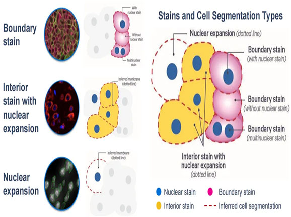
For spatial transcriptomics data analysis, accurate cell boundary segmentation is crucial for identifying cell types and studying cell-cell interactions. Compared to Visium HD, Xenium's newly introduced specialized dye combinations enable precise multimodal cell segmentation, enhancing data accuracy and spatial resolution.

From the above comparisons, it is evident that bothVisium HD and Xenium achieve single-cell level high resolution and are compatible with FF and FFPE samples. However, these two platforms have distinct differencesin their methodologies and applications.
In practical applications, Visium HD and Xenium can be used independently or in combination. For new projects, researchers can first usesingle-cell and/or spatial transcriptomics sequencingto identify genes of interest, then validate them with Xenium for higher precision. If key genes have already been identified,Xenium’s pre-designed probe panels or custom-targeted probes can be used directly for validation.
Omics Empower is a 10x Genomics Certified Service Provider. If you are interested in how our services can support your research, please reach out to our team. Whether you need assistance with sample preparation,single cell sequencing, spatial transcriptomics sequencing, or data analysis, we can handle every step of the process to ensure high-quality results. Contact us today for a consultation!
*All images in this article are sourced from 10x Genomics official materials.
Singapore Global Headquarters: 112 ROBINSON ROAD #03-01
Germany: Kreuzstr. 60, 40210 Düsseldorf
United States: 2 Goddard, Irvine, CA 92618
Singapore Global Headquarters: 112 ROBINSON ROAD #03-01
Germany: Kreuzstr. 60, 40210 Düsseldorf
United States: 2 Goddard, Irvine, CA 92618